Vivek Gopalakrishnan
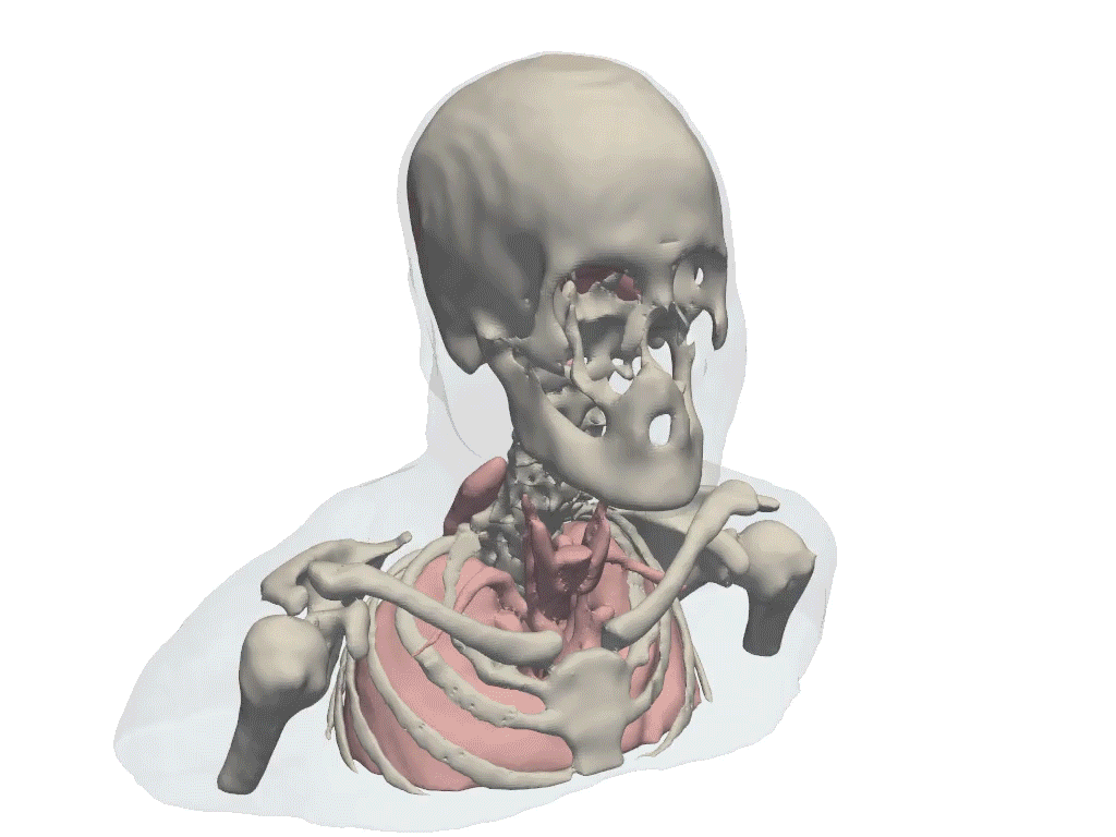
Example visualization from PolyPose, a tool developed by Vivek Gopalakrishnan and colleagues that infers the 3D location of anatomical structures from 2D X-ray images, enabling doctors and surgical robots to navigate without 3D imaging.
GRAPHIC: COURTESY OF VIVEK GOPALAKRISHNAN
Vivek Gopalakrishnan, a PhD candidate in medical engineering and medical physics in the Harvard–MIT Program in Health Sciences and Technology (HST), draws on video game design to create 3D models that help doctors perform delicate brain, spine, and heart procedures.
“People may not know that a lot of what we do is inspired by computer graphics,” says Gopalakrishnan, who uses computational modeling to advance new approaches for minimally invasive, image-guided surgery.
“A lot of the research into making animated films or video games look realistic, especially the math behind rendering 3D scenes as 2D images, is closely related to medical imaging modalities like X-rays,” he says.
“A number of the foundational textbooks I read are ones that Pixar engineers might also read. There are some cool crossovers that don’t seem obvious at first, but when you put the fields together, you arrive at some very powerful solutions.
“You never know where you might draw inspiration from to solve hard problems,” he says.
Creating 3D models from 2D X-rays to better orient surgeons
Gopalakrishnan’s research centers on patient-specific machine learning, developing personalized models that leverage the physics behind X-rays to extract 3D information from 2D imaging.
“Imagine minimally invasive heart procedures,” he says. “Instead of opening the chest and touching the heart, physicians use small tubes to navigate medical devices along the highway of blood vessels to reach the diseased vessel or valve. This approach reduces trauma and recovery time but is very technically demanding.”
“You’re trying to intervene on a moving, three-dimensional structure,” he continues, “but you’re looking at grainy two-dimensional X-rays where all the anatomy is compressed into a single plane. Even after a decade of training, mentally reconstructing the underlying 3D anatomy in real time can still be really hard, especially for challenging cases.”
Gopalakrishnan’s work aims to reduce this cognitive burden. By generating personalized 3D visualizations from 2D images acquired during surgery, his models can help doctors better understand where they are and how to position devices safely—without having to do all the spatial reasoning in their heads.
He is also interested in surgical robotic applications and actively collaborates with startups to deploy his algorithms on robotic systems. “Telling robots where they are in 3D from intraoperative images is the first step towards autonomous navigation and interventions.”
“Taking things clinicians find challenging and making them easier”
Gopalakrishnan earned his bachelor’s and master’s degrees in biomedical engineering at Johns Hopkins University. At MIT, fellowship support has played an important role in shaping his graduate experience. He was a Takeda Fellow in 2023–2024 and currently holds a graduate fellowship through the MIT Health and Life Sciences Collaborative (MIT HEALS). “Being a member of the HEALS community connected me with other graduate students at MIT working on fascinating biomedical problems whom I wouldn’t have met otherwise,” he says.
Advised by Polina Golland, the Sunlin (1966) and Priscilla Chou Professor in the Department of Electrical Engineering and Computer Science and at the MIT Institute for Medical Science and Technology, Gopalakrishnan is affiliated with the Computer Science and Artificial Intelligence Laboratory at MIT.
As a fifth-year doctoral student in the Harvard–MIT HST program—which celebrated its 55th year in 2025—he is among a cohort of clinician-scientists and engineers learning to harness the combined power of science, engineering, and medicine to translate research findings into clinical practice, and to improve human health.
In January, he did a one-month rotation at Mass General Hospital as part of the HST PhD curriculum. “Spending time at MGH has been such a privilege,” he says. “Talking to clinicians, nurses, technologists, and especially patients about the things they find challenging, and then doing research to make those things easier, is a really rewarding goal to work towards.
“Biomedical problems are particularly fun to work on. At MIT, I get to develop elegant mathematical formulations and algorithms that solve the abstract problem on paper, which I find intrinsically enjoyable. Then, I get to go to one of the hospitals across the river, run my code on clinical data, and wrestle with all the messy edge cases until it actually works on real patients. I love constantly iterating between the theoretical and the practical while working on these hard problems.”
TWO WAYS TO SUPPORT EXCEPTIONAL RESEARCHERS
Gopalakrishnan is receiving support through the MIT HEALS Graduate Fellowships, a highly selective fellowship program supporting exceptional graduate students for an academic year. A doctoral student in HST, he brings interdisciplinary training to his work at the intersection of engineering and medicine. You can make your gift to the MIT HEALS Graduate Fellowships or contribute to the HST Founders’ Fund, strengthening the foundation that allows exceptional medical engineers to pursue bold ideas with the rigor and freedom required to translate discovery into real-world impact.