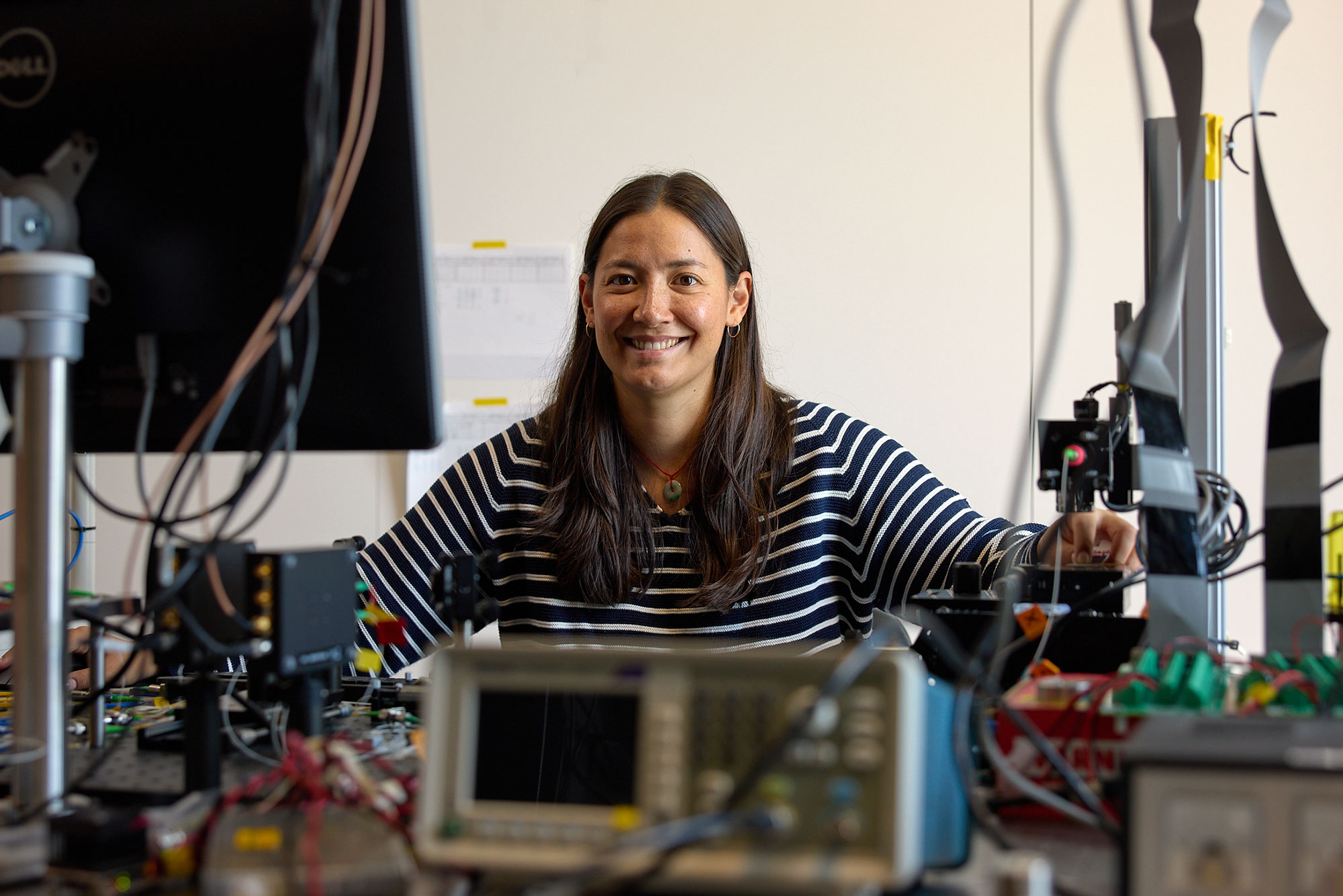
PhD candidate Ginger Schmidt is designing optical medical imaging tools. Here she works at an optical table aligning the reference arm of an optical coherence tomography system.
PHOTOS: KEN RICHARDSON
But the big moment that helped point her toward a medical engineering career occurred when the device made its debut in a physician’s hands.
Although the experimental surgery she witnessed did not involve a real-life patient, Schmidt says the experience brought her whole vision into focus.
“Seeing a device that I helped design perform a surgical procedure was incredibly exciting and rewarding,” says Schmidt, a fifth-year PhD candidate in medical engineering and medical physics in the Harvard–MIT Health Sciences and Technology program. “It transformed my understanding of engineering from a practice that existed in the lab or on a computer screen into a field that could directly impact patient care.”
A fellowship for boundary-crossing science
Today, Schmidt, who received a graduate fellowship through the MIT Health and Life Sciences Collaborative (MIT HEALS), is bringing that same sense of purpose to exploring the use of optics to image heart disease. Her research, which she conducts at Massachusetts General Hospital and Harvard Medical School, brings together optics, physics, signal processing, hardware engineering, biology, and clinical medicine—the kind of boundary-crossing work that MIT HEALS was created to encourage.
Schmidt’s fellowship is funded by Noubar Afeyan PhD ’87, member of the Executive Committee of the MIT Corporation and founder and CEO of Flagship Pioneering. The fellowship supports graduate students who are bold, cross-disciplinary thinkers tackling major challenges in health and life sciences.

Since the 2025 launch of the HEALS Graduate Fellowship, 63 graduate students have received fellowship funding.
After receiving accolades as the first female captain to win the popular robot competition BattleBots, just as she entered MIT in 2021, Schmidt is now applying the same innovation mindset to biomedical imaging. “I like that I can work in fundamental physics, but then I can also build a tangible product, such as an imaging device. And to top it off, I can also take that imaging device and go into the clinic to treat a patient,” she says. “That’s the trifecta of everything I would ever want to work on.”
Using light to detect disease and save lives
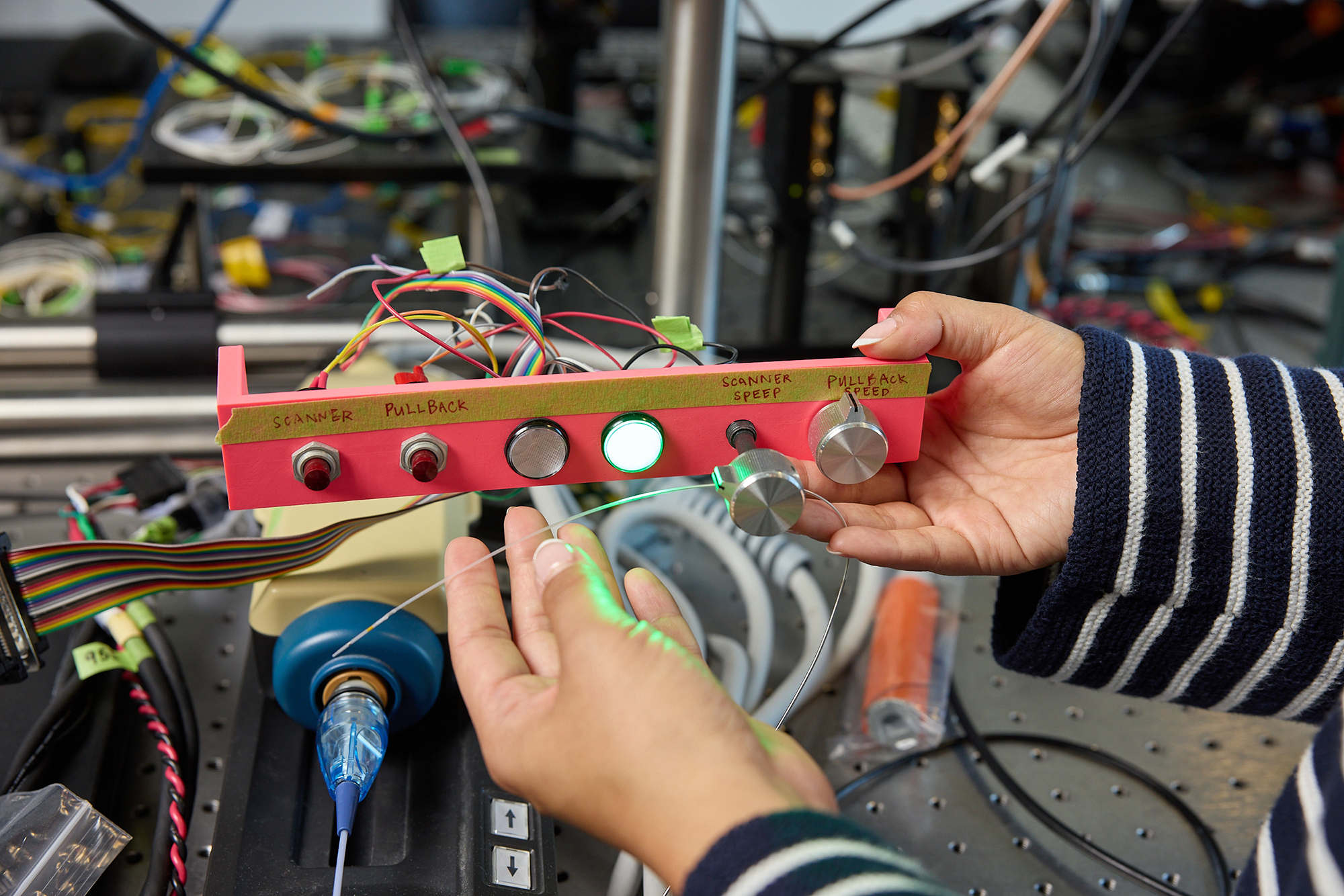
Her research largely focuses on optical coherence tomography (OCT). Schmidt describes OCT as similar to ultrasound, except it uses light instead of sound to form high-resolution, cross-sectional images by measuring light scattered back from tissue.
In eye care, OCT is used to image the retina and cornea. In cardiology, it can be used inside arteries: When a coronary artery becomes blocked, doctors may place a stent, a small mesh tube, to reopen the vessel and restore blood flow. OCT can help physicians check whether the stent is placed correctly, fully expanded, and long enough for the diseased section of the artery.
Schmidt’s research asks whether OCT can do more. Some plaques inside arteries are soft and more likely to rupture, increasing the risk of a heart attack. Others, however, are hardened by calcium and may require additional preparation before a stent can be safely and effectively implanted.
“My PhD research aims to expand what OCT can tell us by measuring how stiff or soft the artery is, not just what it looks like,” she says. “This information could help physicians make more informed treatment decisions.”
Making advanced imaging more scalable
Elastography is the general term for a technique for measuring tissue stiffness. But Schmidt’s goal is not simply to add a new measurement method; it is to create a technology that can be widely and affordably used for patients without major changes to existing equipment or workflows.
Researchers often face a temptation to solve hard medical imaging problems with faster, more powerful, more expensive, or more specialized tools and systems, she says. “It’s exciting to be the first person to see a new biological phenomenon with a novel imaging system,” she says. “But if that system costs more than $500,000 and takes hours to acquire a single image, it’s unlikely to ever make it into routine patient care.”
Schmidt is taking a different route, using signal processing and software. The technique is roughly 50 times faster than many existing approaches and is compatible with current commercial imaging systems, she says. It also does not require specialized scan patterns, ultrafast lasers, or other expensive custom hardware, meaning it can be readily deployed to clinics around the world.
“My hope is that this work will eventually improve patient care and, one day, lead to an FDA-approved technology that can be used routinely in the clinic,” Schmidt says. Some of her findings were published in the Journal of Biomedical Optics in 2025.
Backing research at a pivotal moment
The HEALS fellowship has supported that work at a difficult time for biomedical research amid funding uncertainties.
“The fellowship has been instrumental in supporting both my research and my lab community,” Schmidt says. “It’s given me the freedom to pursue research I am passionate about, continue my education during my PhD, and helped provide stability for my lab during a particularly difficult time.”
She says the support has also helped her lab preserve positions for postdoctoral researchers. And it gives her room to pursue work across fields. For instance, researchers are increasingly growing three-dimensional tissue models to study biology in ways that more closely resemble living tissue.
Many traditional microscopy techniques work best on thin, transparent tissue sections, which involves slicing samples. OCT, by contrast, can image deeper into thicker tissue without destroying samples. Although it does not achieve the same resolution as many microscopy techniques, this trade-off could make it useful for more quickly screening many more drugs to identify treatments that might help treat or cure disease, Schmidt says.
Looking ahead
The daughter of two software engineers, Schmidt jokes that she “rebelled” by going into the more hands-on fields of electrical and mechanical engineering, and then biomedical engineering. Imaging devices are especially appealing to her because a prototype can be built, refined, and used to image tissue or patients on a shorter timeline than drug development or therapeutics require.
For Schmidt, who hopes to become a professor at an engineering school, with a dual appointment at an academic research hospital, that direct line between the lab and the clinic continues to motivate her.
“At places like MIT, we develop remarkable new tools and therapies, but there can be a significant gap between what is technologically possible and what patients are able to access,” she says. “To me, the true measure of success is not whether we can invent a new technology, but whether we can ensure that the people who need it are actually able to benefit from it.”
SUPPORT GRADUATE FELLOWSHIPS IN HEALTH AND LIFE SCIENCES
Ginger Schmidt is receiving support through the MIT HEALS Graduate Fellowships, a highly selective fellowship program supporting exceptional graduate students for an academic year. A doctoral student in the Harvard–MIT Health Sciences and Technology program (HST), she brings interdisciplinary training to her work at the intersection of engineering and medicine. You can make your gift to the MIT HEALS Graduate Fellowships or contribute to the HST Founders’ Fund, strengthening the foundation that allows exceptional medical engineers to pursue bold ideas with the rigor and freedom required to translate discovery into real-world impact.